Skintell Mole Guide

A comprehensive resource for identifying mole types and recognising the signs of skin change.

Seborrhoeic Keratosis

These below are seborrhoeic keratoses. They are usually greyish brown and scaly. The surface is rougher than a mole and they can be friable. They are most often found on the torso but can also be on other sites. They appear from the age of 30 onwards. They usually increase in numbers with age and are not related to sun exposure. They never transform into skin cancer.

Types of moles (naevi)

This shows different types of naevi: flat (junctional), compound (mixed) and intradermal (fleshy)

keychain2 - Moles & Naevi

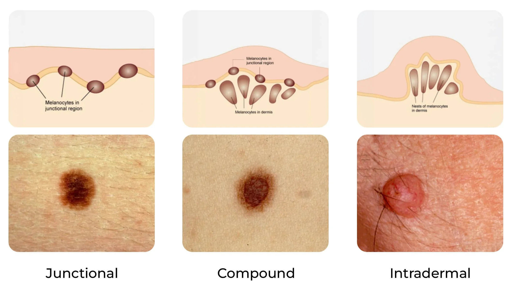
Moles/naevi/atypical moles

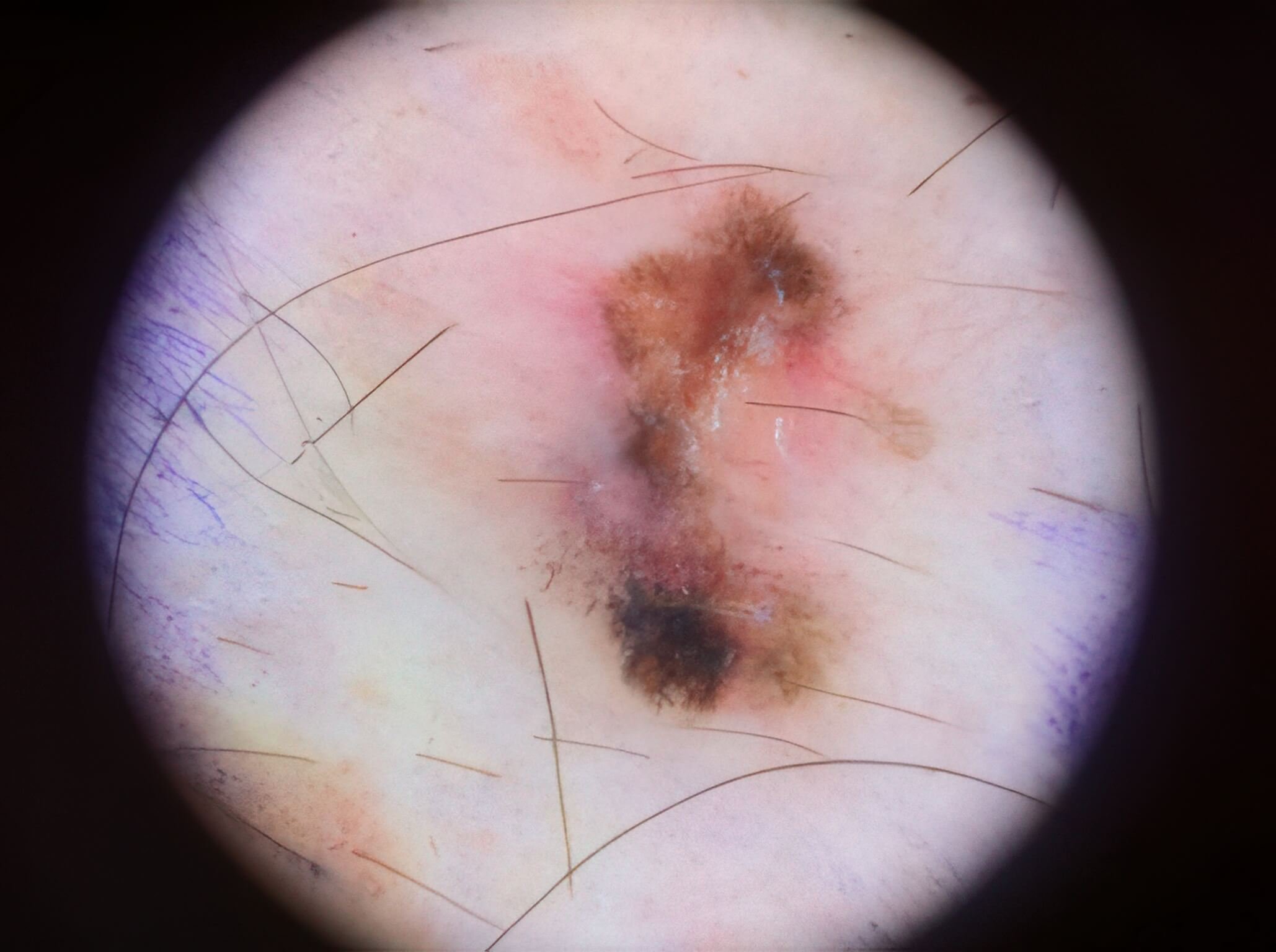
These pictures show multiple atypical naevi ( usually above 5 mm in diameter). When these atypical naevi are found in large numbers, it is called the atypical moles syndrome phenotype. This phenotype increases the risk of melanoma and mole mapping every year is important to look for changes over time. People with the atypical mole syndrome may have a family history of melanoma and/or multiple cancers so this needs to be documented so the risk of melanoma can be assessed more accurately.

Naevi (moles) appear in childhood and increase in number and size throughout early adulthood. After the age of 40 years, it is unusual to still acquire new naevi unless one belongs to a very moley family. Melanoma is extremely are in children and they are not screened for melanoma below the age of 18. There are exceptions for children with very large birthmarks covering large body areas.

The chance of transformation into melanoma for each mole is very low but the presence of multiple large naevi is a marker of increased melanoma risk. Prophylactic excisions of moles is not the right way to manage people at high risk as it is important to note that a melanoma may develop from normal skin with no mole present before.

Dermatofibroma

These are dermatofibromas. These benign skin lesions are often found on the legs especially in women but can be found in males and other sites of the body. They usually have a paler centre with a darker rim of brown all around the lesion. They are firm and when the lesion is squeezed, a dimple is seen which confirms that the lesion is deep in the dermis as it comes from the collagen layer of the skin. They never progress to skin cancer.

Haemangioma

These show haemangiomas which are benign. They are formed of blood vessels and are more purplish red than melanoma. They have a soft surface but may bleed heavily if they are traumatised. Some haemangiomas may be difficult to differentiate from a non pigmented melanoma and dermoscopy ( 20 magnification) is important to diagnose these benign lesions.

Basal Cell Carcinoma (BCC)

This is a basal cell carcinoma with pigmentation. The edges are often raised and shiny. Basal cell carcinomas grow very slowly over months and are not dangerous as they never spread. They are usually found in older individuals above 50 years of age. But they can also be seen in younger individuals especially on the torso when they can be more superficial. They can be treated with surgery, creams, cryotherapy (freezing) or photodynamic therapy depending on the site and size.

Squamous Cell Carcinoma (SCC)

These are squamous cell carcinomas. They present as a raised nodule and most often with a crust and found on chronically sun exposed areas. They are very rare before the age of 50. They are found on very damaged sun damaged skin. They grow faster than basal cell carcinoma. They are usually easily treatable with surgery but, in rare cases, can spread to the lymph nodes. When they are very crusty, they grow slowly and very unlikely to spread to other parts of the body. When they have no crust and are very red, they may bleed on contact. Those are fast growing and may spread. However, the latter are rare and most people with squamous cell carcinoma do well with surgery alone. 

Early Melanoma

These are early melanomas as they have irregular borders and different shades of colour. Black, blue, white and red are regarded as suspicious in a mole. All shades of brown are allowed but the colour should be fairly symmetrical. If you try to cut the mole into 4 quadrants the parts do not longer match in melanoma so loss of symmetry is a very important sign to look for. Most melanomas have dark colours including black but, rarely, in people with fair skin, they can present as a red nodule without dark pigment. Melanomas grow in weeks and months. Most melanoma are treated with surgery alone but around 10% can spread to the lymph nodes. They are the most dangerous form of skin cancer but luckily the rarest. The mean age for melanoma in the UK is 55 years of age. A high number of moles especially large ones is the most important risk factor for melanoma. Family history of melanoma also raises the risk. Fair hair and skin with a lot of sun exposure are risk factors too.

guide9 - Advanced Melanomas

Advanced Melanomas

Melanomas with regression showing white colours because the immune system has attacked the tumour.